Preparing the blood sample for KHV Serological testing ..
The virus neutralization test is done with a very small amout of serum or plasma from the fish being tested. As the sample will be grown in tissue culture with Koi Fin cells and KHV, we wish to avoid contaminating the sample durning handling and separation of the serum or plasma. From a practical standpoint, it is quick and economical to transfer the blood to a vacutainer after it is drawn and before it clots in the syringe.
Transferring the sample..
Tranferring the blood to the microtainer involves puncturing the top of the microtainer with the syringe needle. As the microtainer is under a vacumn, the plunger of the syringe should be initally held in ordeer to retart the inital blood flow. Additionally, it is helpful to hold the needle against the inside wall of the tube. These two measures aid in reducing hemolysis (the lysis of the fragil red blood cells). Again, one does not forcefully shoot the blood from the syringe but rather inhits its progress into the vacutainer, holding the side of the needle against the wall to prevent "splashing".
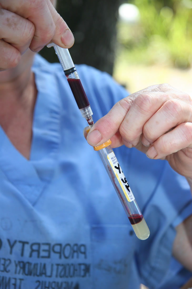
If the volume of blood is so small that microtainers must be utilized, care must be taken not to contaminate the inside of the tube or the inside of the cap when the blood is transferred. Microtainers are not sterile and the manufacturer indicates they should not be sterilized, although they are "clean" from the bag and seem to work well as long as contamination is avoided. Prompt refrigeration after spinning retarts possible bacterial or fungal contamination growth.
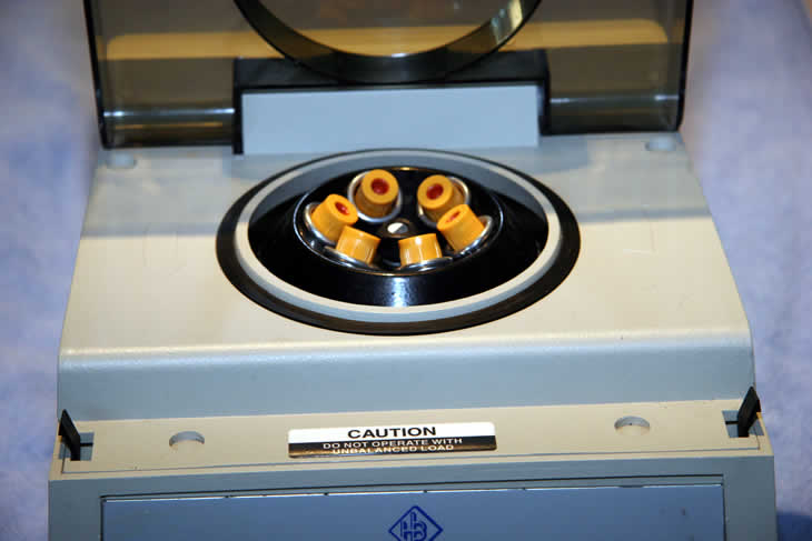
Centrifugation...
Specimens should have the red cells separated from the serum or plasma as soon as pratical. Once the serum or plasma is separated from the red cells, the specimen is much more stable. To separate the red cells from the plasma or serum, we use a centrifuge. It usually to takes ten to fifteen minutes of centrifugation time to obtain adequate separation.
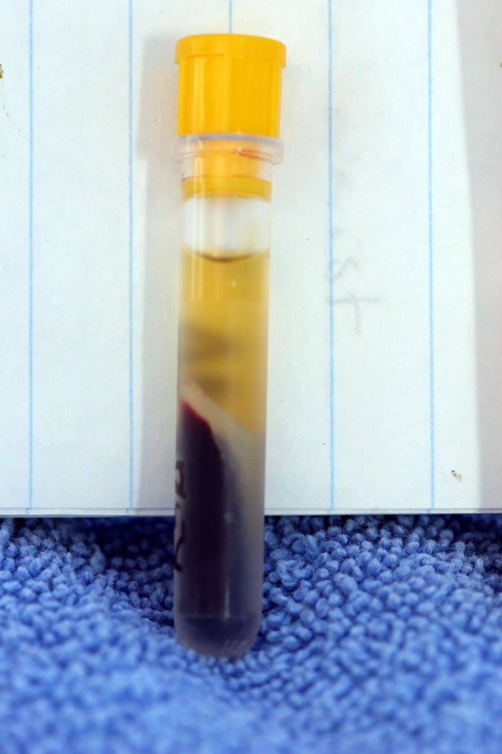
Following centrifugation, the red cell mass is at the bottom with the gel layer separating the red cells from the serum or plasma as shown in the photos. The samples are kept in the refigarator if shipping will be done over the next day or two. At this point the specimen can be frozen if samples will be collected over several days before shipping. Repeated thawing and freezing is to be avoided.
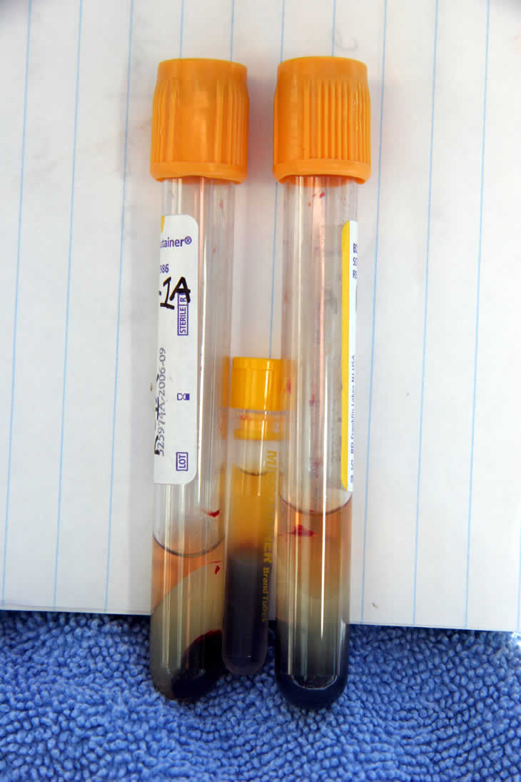
Vacutainers on either side of microtainer after spinning. Serum on top, gel in center layer and red cell mass at base.
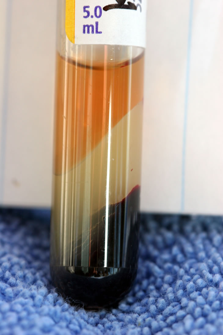
Typical sample in a vacutainer after centrifugation.
Microtainer with sample after centrifugation. A lessor amount of blood could have been utilized.
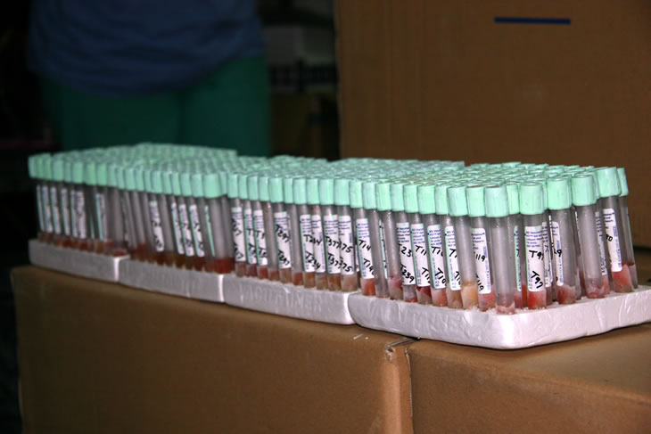
A group of samples collected over three days, frozen after centrifigation,and awaiting shipment to the laboratory.
One fish, one sample, one test!
Samples for KHV serological testing should not be combined!
How to Accidentally Alter or Contaminate a Sample so as to make it Worthless for a KHV virus neutralization assay (and give you an erronous result).